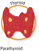
Thyroid Nodules
 Thyroid nodules are abnormal growth of thyroid cells which form a nodule in the thyroid gland. Often these abnormal growths of thyroid tissue can be felt as a lump in the thyroid and can even sometimes be seen as a lump in the front of the neck. Some thyroid nodules may produce too much thyroid hormone (hyperthyroidism). However, most nodules do not produce thyroid hormone but can cause symptoms such as neck pain, difficulty in swallowing, shortness of breath or change in the voice. Thyroid nodules are very common:
• 7-8% of young women have a thyroid nodule
• 2-3% of young men have a thyroid nodule
• Most people will develop a thyroid nodule by the time they are 50 years old and risk increases with age:
o 50% of 50 year olds will have a least one thyroid nodule
o 60% of 60 year olds will have a least one thyroid nodule
o 70% of 70 year olds will have a least one thyroid nodule
• More than 90% of all thyroid nodules are benign (non-cancerous growths) and 5-10% are cancerous
• Some are actually simple cysts, which are filled with fluid rather than thyroid tissue
We do not know what causes most non-cancerous thyroid nodules. Sometimes inflammation (Hashimoto’s) of the thyroid causes nodules and rarely iodine deficiency can do it, but most nodules appear to be a genetic defect.
These nodules are usually diagnosed after a patient complains of symptoms or are found during a routine physical exam. Sometimes they are found on screening tests done for other reasons, such as an x-ray, ultrasound or CT scan done of the neck. Once a nodule is diagnosed, a routine history and physical exam should be done by a physician with expertise in this area. Your physician will feel the thyroid to see whether the entire gland is enlarged, whether there is a single nodule present, or whether there are many nodules in your thyroid. The initial laboratory tests may include blood tests to measure the amount of thyroid hormone (Thyroxine or T4) and thyroid-stimulating hormone (TSH) in your blood to determine whether your thyroid is functioning normally. Most patients with thyroid nodules will also have normal thyroid function tests. Some tests may also include TPO (an antibody which destroys the thyroid) to characterize the cause of why some people get thyroid nodules.
A thyroid ultrasound is also commonly ordered to determine the size, location and sometimes the composition of the nodule(s).
The thyroid ultrasound is usually done as the first test and a biopsy FNA (fine needle aspirate) of the nodule helps to characterize the cells in the nodule. However, a scan is usually ordered if an overactive thyroid is suspected.
A biopsy of the nodule with a fine needle aspiration (FNA) will tell if the nodule is cancerous or benign. A fine needle biopsy of the thyroid nodule may sound frightening, but the needle used is very small and a local anesthetic is usually not needed. This simple procedure is done in the doctor’s office. It does not require any special preparation (no fasting), and patients usually return home or to work after the biopsy without any ill effects. For a fine needle biopsy, the doctor will use a very thin needle to withdraw cells from the thyroid nodule. Ordinarily, several samples will be taken from different parts of the nodule to give the doctor the best chance of finding cells to be evaluated by a pathologist to determine the nature of the nodule. Most frequently a thyroid ultrasound can be used to guide the needle directly into the nodule. The cells are then examined under a microscope by a pathologist.
The report of a thyroid fine needle biopsy will usually indicate one of the following findings:
1. The nodule is benign (noncancerous). This result is obtained in 50% to 60% of biopsies and the risk of overlooking a cancer when the biopsy is benign is generally under 3%. Generally, these nodules need not be removed, but another biopsy may be required in the future, especially if they get bigger.
2. The nodule is malignant (cancerous). This result is obtained in about 5% of biopsies and often indicates papillary cancer, one of the most common thyroid cancers. All cancerous nodules should be removed surgically, preferable by an experienced thyroid surgeon.
3. The nodule is suspicious. This result is obtained in about 10% of biopsies and suggests that the nodule may be a cancer. These nodules need to be removed surgically to determine if the nodule is or is not cancerous.
4. The biopsy is non-diagnostic or inadequate. This result is obtained in up to 20% of biopsies and indicates that not enough cells were obtained to make a diagnosis. This is a common result if the nodule is a cyst. These nodules may be removed surgically or, more often, be re-evaluated with second fine needle biopsy, depending on the clinical judgment of your doctor.
A thyroid scan is sometimes needed and uses a small amount of a radioactive substance, usually radioactive iodine, to obtain a picture of the thyroid gland. Because thyroid cancer cells do not take up radioactive iodine as easily as normal thyroid cells do, this test is used to determine the function of the nodule or the likelihood that a thyroid nodule contains a cancer. If done as the first test, the thyroid scan is used to determine those patients who most need a biopsy. The scan usually gives the following results:
1. The nodule is “cold” and non-functioning. In other words, the nodule is not taking up radioactive iodine normally. This patient is referred for a fine needle biopsy of the nodule.
2. The nodule is “warm” and functioning. Its uptake of radioactive iodine is similar to that of normal cells. A biopsy is usually performed but the likelihood of cancer is lower than a “cold” nodule.
3. The nodule is hot. Its uptake of radioactive iodine is greater than that of normal cells and suggests the nodule is overproducing thyroid hormone. The likelihood of cancer is extremely rare, and so biopsy is usually not necessary.
![]()


